
Light sheet fluorescence microscopy (LSFM) is an imaging method that uses one or more illumination objectives to excite fluorophores in a specimen with a thin sheet of light, which are then visualized with a separate imaging objective perpendicular to the sheet. LSFM has several advantages over confocal microscopy, including:
- Excellent sample penetration
- High speed
- Minimal phototoxicity and photobleaching
These properties make LSFM ideal for many biological imaging applications, including:
- Fluorescence imaging of thick samples
- Long term imaging of live samples
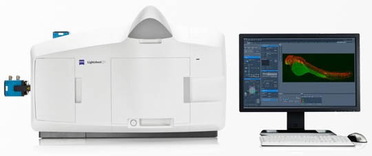 Image courtesy of Carl Zeiss AG
The Zeiss Lightsheet Z.1 includes several additional features to optimize LSFM:
- Four-axis sample positioning permits rotation of the specimen for easy viewing from any angle.
- Multiview acquisition allows the same tissue to be imaged from multiple perspectives, then integrates these views for optimal resolution and accurate rendering.
- Two illumination objectives, one on each side of the sample chamber, enhance even illumination of the specimen.
- Pivot scanning slightly rotates the sample during acquisition to eliminate shadowing and striping artifacts observed in other systems
- Cleared tissue optics (5x objective) designed for CLARITY and similar clearing techniques (RI=1.45)
For more information about LSFM imaging methods, visit MicroscopyU and for details about the Lightsheet Z.1, visit the Carl Zeiss Microscopy. To discuss whether light sheet microscopy would be of benefit for your application, please contact us for a free consultation.
System specifications:
The Zeiss LightSheet Z.1 Dual Illumination Microscope System is ideally suited for imaging of thick and/or live specimens, because it uses separate illumination and detection light paths to minimize photobleaching and phototoxicity and maximize imaging depth. The 4-axis sample positioning system allows imaging from any perspective and Multiview acquisitions increase axial resolution. The system features two illumination objectives and uses ultrafast pivot scanning to reduce shadowing artifacts. The system includes temperature and CO2 environmental control of the sample chamber. Solid-state lasers provide excitation lines at 405, 445, 488, 515, 561, and 638nm, with simultaneous two channel detection using PCO.edge 16 bit sCMOS cameras. Imaging objectives include 5x dry and 20x and 40x water immersion lenses, with 2.5x system optical zoom, yielding effective magnifications of 2x-100x. Optics for cleared tissue imaging using a 5x objective are designed for a refractive index of 1.45, matching that used in CLARITY and similar techniques. Image acquisition and basic manipulation is performed with Zen software, that also integrates with Arivis Vision 4D for rendering and analysis. A separate workstation is available for these analyses.
